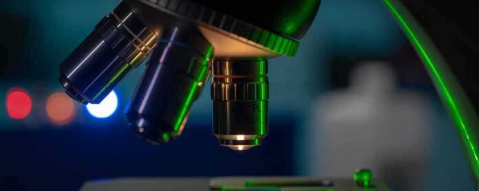
Scientists at Stanford can now watch the inside of a living cell move and change in real time, without injecting it with a single glowing dye.
Quick Take
- Stanford chemists built a new microscope called Interferometric Image Scanning Microscopy, or iISM, that images live cells without fluorescent labels.
- The tool reaches about 120 nanometers of lateral resolution, sharp enough to track tiny structures moving inside a living cell.
- It uses less laser light than older methods, letting researchers watch cells longer without damaging them.
- The findings appeared in the peer-reviewed journal Light: Science & Applications in 2026, though Stanford itself calls the device a prototype.
A Microscope Built To See Cells On Their Own Terms
Researchers led by W. E. Moerner at Stanford’s Chemistry Department developed a microscope that blends two existing techniques: image scanning microscopy and interferometric detection. Instead of relying on fluorescent tags to light up cell parts, iISM reads the light that naturally scatters off structures inside a cell. A computer then reconstructs that scattered light into a detailed, real-time picture, according to Stanford’s own research description.
That label-free approach matters because fluorescent dyes, while useful, can change how a cell behaves or wear it down under bright light. Stanford’s team reports that iISM lets scientists see internal structures interacting as they naturally move, without forcing the cell to carry an artificial tag just so it can be seen.
Why 120 Nanometers Is A Big Deal
The peer-reviewed paper describing the work, published in Light: Science & Applications, documents label-free imaging inside live cells at roughly 120 nanometers of lateral resolution. For comparison, a human hair is about 80,000 to 100,000 nanometers thick. That kind of resolution lets scientists watch details far smaller than what a standard light microscope could ever pick up, all while the cell stays alive and undisturbed.
Stanford describes this as the highest resolution achieved so far without fluorescent labels, a claim that stands out because label-free imaging has historically traded away sharpness for safety. Getting both at once is the technical leap researchers have chased for years.
Gentler Light, Longer Observation Windows
One of the microscope’s most practical selling points is reduced light exposure. Stanford’s technology description states that iISM keeps its imaging speed while using less laser power than comparable systems, which lowers the risk of damaging the very cells researchers are trying to study. That matters because scientific literature has long documented a tradeoff in super-resolution imaging: sharper pictures usually demand brighter light, and brighter light can injure or kill the sample being studied.
Reviews of super-resolution microscopy describe this balancing act as a persistent four-way compromise between resolution, speed, image clarity, and cell health. Label-free techniques like iISM are part of a broader scientific push to sidestep that tradeoff by cutting down on how much light a cell has to absorb just to be photographed clearly.
Stanford Calls It A Prototype, Not A Finished Product
Despite the strong results, Stanford’s own technology-transfer listing classifies iISM as a prototype, or proof of principle. That label matters. It signals the instrument has proven the concept works but has not yet been mass-produced, standardized, or tested across the wide range of cell types and labs where future scientists might want to use it.
Stanford also frames iISM as complementary to fluorescence microscopy rather than a replacement for it. Fluorescent labeling remains the gold standard for identifying specific molecules inside a cell, something scattered-light imaging alone cannot yet match. iISM’s strength is showing structure and movement broadly, not pinpointing individual molecules by name.
What Comes Next For This Technology
Researchers have floated potential uses ranging from studying how pathogens invade cells to tracking drug delivery, plant-microbe interactions, and cancer biology. Those applications remain forward-looking rather than completed studies. The next real test for iISM will be whether other labs outside Stanford can replicate its resolution and gentle-light performance on their own equipment and their own cells.
For now, the achievement stands on its own: a working microscope that lets scientists watch living cells behave naturally, in sharp detail, without forcing them to wear a tag first. That is a meaningful step forward for a field that has spent decades trying to see more without breaking what it’s looking at.
Sources:
sciencedaily.com, newsy-today.com