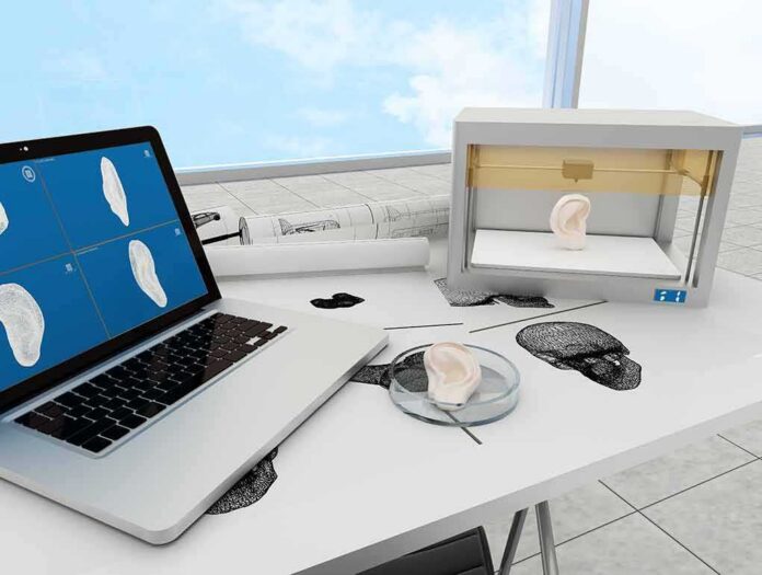
Mayo Clinic researchers have built a living, 3D-printed human skin model that survives for weeks in the lab — and the Food and Drug Administration (FDA) is already reviewing it as a potential replacement for animal testing.
Story Snapshot
- Mayo Clinic created the first fully humanized 3D bioprinted skin model using plant-based collagen — no animal materials involved.
- The printed skin mirrors real human skin layers and can stay alive for weeks, far longer than traditional lab samples.
- The FDA is actively reviewing the technology as a possible alternative to large animal studies in drug testing.
- Blood vessels, nerves, and immune cells are still missing from the model — key hurdles before it reaches patients.
What Mayo Clinic Actually Built and Why It Matters
Researchers at Mayo Clinic partnered with biotech firm CollPlant to create a skin model printed with recombinant human collagen — a plant-grown protein that mimics the collagen in your body. That matters because most lab-grown tissue models rely on animal-derived materials, which can trigger immune reactions and produce inconsistent results. This model uses entirely human-derived components, cutting out that variability at the source. The result looks and behaves like real skin at the cellular level.
The printed tissue replicates skin’s layered structure, including pigment-producing cells called melanocytes. Side-by-side comparisons of the printed model and actual patient skin show nearly identical cellular architecture and biological responses. That kind of structural accuracy is what separates a true research tool from a novelty. Dermatopathologists reviewed the printed tissue in blinded tests to confirm it matched real human skin samples — a rigorous standard most lab models never reach.
The Weeks-Long Survival Window Changes Everything for Researchers
Traditional skin explants — small pieces of real skin kept alive in a dish — die within days. The Mayo Clinic bioprinted model survives for weeks. That extra time lets scientists watch how diseases progress slowly, how wounds heal, and how a patient’s skin responds to a drug over a realistic timeline. Think of it like the difference between a snapshot and a full-length film. Chronic wound research, skin aging studies, and drug testing all become far more meaningful with that extended window.
Because the model is printed, it can be reproduced with remarkable consistency — the same structure, the same cell types, the same layering every time. That reproducibility is critical for drug testing. If your test platform varies batch to batch, your results do too. Printing solves that problem by turning a biological process into something closer to manufacturing. Researchers can also tailor the model to reflect a specific patient’s biology, printing skin based on that individual’s own cells.
FDA Engagement Signals a Real Shift in Preclinical Testing Standards
The FDA reviewing this technology is not a formality. The agency has historically required extensive animal studies before human trials. The fact that it is now engaging with Mayo Clinic’s team — examining data on how well printed skin replicates human tissue structure and function — suggests regulators see genuine potential here. That does not mean approval is imminent. Regulatory clearance for novel tissue-engineered products moves slowly, and the complexity of validating these models for routine use remains a real barrier.
What the Model Still Cannot Do
The current prototype has no blood vessels, no immune cells, and no nerve fibers. Those are not minor omissions. Blood vessels deliver oxygen and nutrients to real skin. Immune cells drive inflammation and healing. Nerves control sensation. Without them, the model cannot fully replicate how skin responds to infection, injury, or immune-mediated diseases like psoriasis or eczema. The Mayo Clinic team openly acknowledges these gaps and says future versions will work to include them. That honesty is refreshing — and it sets realistic expectations for what this technology can do right now versus what it might do in five years.
Replicating nerve networks is particularly hard. Nerves are not just structural — they signal, adapt, and connect to the central nervous system in ways that a printed scaffold cannot yet mimic. Getting blood vessels to grow into printed tissue is also an unsolved challenge at clinical scale, though animal model studies have shown host vessels can grow into bioprinted constructs after implantation. These are solvable problems, but they will take time and significant research investment to crack.
The Bigger Picture — Animal Testing Is on Notice
Every year, millions of animals are used in preclinical skin research. Many of those studies produce results that do not translate well to humans because mouse or pig skin differs meaningfully from human skin. A validated, fully humanized printed skin model could reduce that gap — and reduce the number of animals needed. That is a practical win for science and a moral one too. The technology is not there yet for routine clinical use, but the direction is clear. What Mayo Clinic has built is not science fiction. It is a working prototype with FDA attention, peer-reviewed publication, and a credible roadmap. The gap between “promising lab model” and “standard clinical tool” is still wide — but for the first time, researchers can actually see across it.
Sources:
youtube.com, prnewswire.com, newsnetwork.mayoclinic.org, mayoclinic.elsevierpure.com